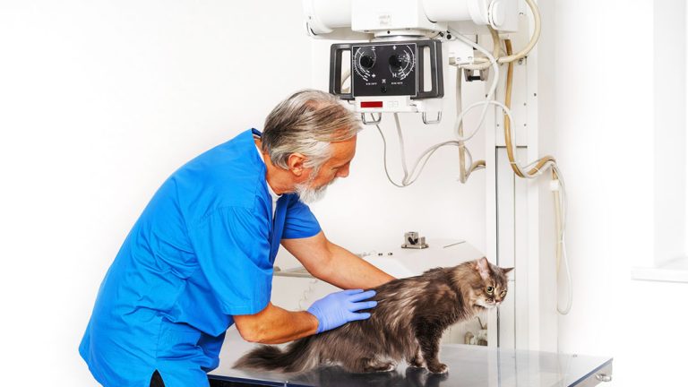
Introduction: The Human Element in Veterinary Radiology
Great veterinary radiology involves more than technology alone—it’s about the people who operate that technology. In our previous blog, “Digital Transformation in Veterinary Radiology Equipment and Workflow Optimization,” we explored the technological advances revolutionizing veterinary imaging. Now, we turn our attention to the equally important human elements that make these technologies truly effective.
While advanced equipment provides the foundation, it’s your team’s expertise, efficient workflows, and clear communication that transform good images into excellent patient care. The most successful radiology practices blend sophisticated equipment with skilled personnel and effective client interactions.
This blog explores how focusing on the human factors—training, resources, and communication—can significantly improve radiographic outcomes, practice efficiency, and client satisfaction.
Building a Skilled Team Through Comprehensive Training
Your team’s proficiency with radiology equipment and protocols sets the stage for everything else. Continuous training enhances individual skills while ensuring smooth, safe, and efficient radiographic processes.
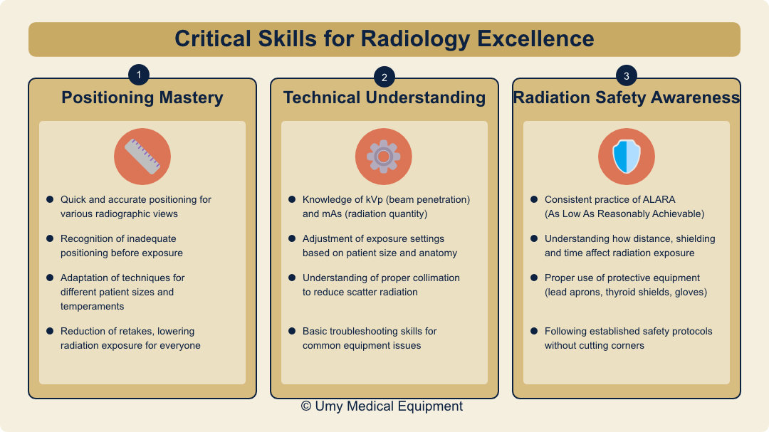
Critical Skills for Radiology Excellence
Positioning Mastery
Proper animal positioning is fundamental to obtaining diagnostic-quality images. Well-trained staff quickly and accurately position animals for various radiographic views and recognize when positioning is inadequate before exposure. They adapt techniques for patients of different sizes, temperaments, and conditions, which reduces the need for retakes and lowers radiation exposure for everyone.
Technical Understanding
Staff should comprehend the technical aspects of your equipment, including the relationship between kVp (beam penetration) and mAs (radiation quantity). Knowing how to adjust exposure settings based on patient size and anatomy makes a significant difference in image quality. Understanding the importance of proper collimation to reduce scatter radiation and basic troubleshooting for common equipment issues saves time and improves results.
Radiation Safety Awareness
Safety remains the top priority in any radiology department. All staff must consistently practice ALARA (As Low As Reasonably Achievable) principles and understand how distance, shielding, and time affect radiation exposure. Proper use of protective equipment (lead aprons, thyroid shields, gloves) and following established safety protocols without cutting corners protect both staff and patients.
Effective Training Methods and Resources
Initial and Refresher Training
Equipment vendor training upon initial purchase or upgrade provides a strong foundation. Structured onboarding programs for new staff ensure everyone starts with the same knowledge base. Annual refresher sessions on safety and best practices keep skills sharp, while cross-training ensures coverage during staff absences.
Hands-On Learning Opportunities
Supervised practice sessions with constructive feedback help staff develop confidence and competence. Peer teaching between experienced and newer staff builds team cohesion while reinforcing knowledge. Quality review of radiographs with discussion of improvement areas helps everyone learn from both successes and mistakes.
External Learning Resources
Several valuable external resources can enhance your team’s radiology skills:
- Professional organizations and courses – Organizations like the Academy of Veterinary Technician Diagnostics Imaging and American Association of Veterinary Radiologists offer specialized continuing education
- Online platforms and webinars – Digital learning environments provide flexible, accessible training options that fit busy clinical schedules
- Peer networks and forums – Connecting with other veterinary professionals allows for case discussions and shared learning experiences
These resources help your team stay current with evolving best practices while building professional connections in the field.
Modern Tools for Enhanced Interpretation
The value of radiography doesn’t end with image capture—accurate interpretation is equally crucial. Today’s veterinarians have access to more interpretation tools and resources than ever before.
Online Databases and Reference Materials
Veterinary professionals can improve their diagnostic skills through various digital resources. Veterinary-specific databases provide comprehensive collections of research and clinical information relevant to radiology. Medical literature databases offer vast repositories of studies applicable to veterinary practice. Digital reference tools allow quick lookups of radiographic findings, while online veterinary manuals provide detailed information about radiographic appearances of various conditions.
Essential Reference Books
Having good reference materials within reach improves confidence and accuracy in interpretation. Atlases of normal radiographic anatomy help distinguish normal variants from pathology—a crucial skill for accurate diagnosis. Comprehensive textbooks on veterinary diagnostic radiology provide detailed references covering principles and interpretation techniques. Species-specific radiographic guides offer focused information on common domestic and exotic animal patients.
AI-Powered Diagnostic Support
Artificial intelligence tools now serve as valuable assistants in radiographic interpretation. AI screening tools help review images rapidly for common conditions, while decision support capabilities focus attention on areas needing closer examination. Many practices use AI-identified findings as teaching tools for staff development.
These tools support—not replace—clinical judgment. Their greatest value comes from reducing decision fatigue through preliminary screening, providing a “second look” that may catch subtle findings, and improving workflow efficiency through faster initial assessment.
Teleradiology Services
When cases become complex, connecting with board-certified radiologists through teleradiology services provides access to specialized knowledge. These services offer rapid interpretations to speed diagnosis and treatment. Many veterinarians use these consultations as learning opportunities for their entire team, reviewing specialist findings to build internal expertise.
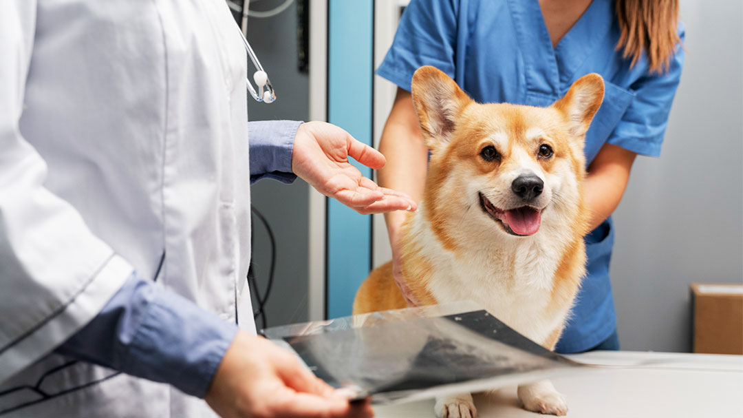
Communicating Effectively with Clients
Even the most accurate radiographic interpretation loses value if clients don’t understand the findings and recommendations. Clear communication builds trust and ensures pet owners understand their pet’s condition and care options.
Explaining Radiographic Findings
When discussing x-ray results with clients, use simple, non-technical language rather than medical jargon. Showing actual images when appropriate and pointing out relevant findings helps clients visualize the problem. Visual aids such as anatomical models can enhance understanding, while clear explanations of what findings mean for their pet’s health provides context.
For example, rather than saying “The radiograph shows pulmonary infiltrates consistent with bronchopneumonia,” consider: “The x-ray shows an infection in your dog’s lungs, which explains the coughing you’ve noticed. Let me show you the cloudy areas that indicate inflammation.”
Discussing Treatment Plans
Transparency and empathy build client trust and compliance. Clearly outline treatment options with their benefits, risks, and costs, and listen to client concerns and address questions patiently. Offering different care options accommodates varying budget constraints, while providing written summaries gives clients reference materials to review at home.
Remember that clients often need time to process information, especially when concerned about their pet. Consider following up the next day for complex cases to answer any additional questions that may have arisen.
Measuring Your Success
Implementing ongoing quality assessment helps identify areas for improvement in your radiology practice.
Key Performance Indicators (KPIs)
To meaningfully assess your radiology performance, track these essential metrics:
- Image quality measurements – Monitor retake rates, reasons for repeated images, and overall diagnostic quality
- Efficiency indicators – Track turnaround times from request to completed image and from image acquisition to interpretation
- Financial metrics – Analyze the cost-effectiveness of your radiology services, including equipment utilization rates
- Client satisfaction scores – Gather targeted feedback about the radiology experience, explanation quality, and overall service
Regular review of these metrics helps identify trends, address issues early, and celebrate improvements in your radiology service.
Regular Team Reviews
Schedule periodic reviews where staff can examine interesting or challenging cases and discuss difficulties and potential solutions. These sessions allow team members to share successful techniques and recognize individual and team achievements, building a culture of continuous improvement.
Final Thoughts: The Human Factor Makes the Difference
Effective veterinary radiology depends equally on skilled people and quality equipment. By investing in training, embracing new resources, and clearly communicating with clients, you create an environment where both animals and their owners feel cared for and respected.
Remember, technology makes your job easier—but it’s your team’s skills, compassion, and clear communication that truly make the difference. In an era of increasingly sophisticated equipment, the human element remains the most crucial component of exceptional radiographic care.